
PanCanAID is a philanthropic initiative that strives to enhance the early detection, diagnosis, and treatment of pancreatic cancer patients. This website provides comprehensive information about the PanCanAID team, its objective, and its ongoing projects. Furthermore, our monthly newsletter highlights the latest advancements in pancreatic cancer management. We are committed to broadening our collaboration with teams and institutes who share our vision of improving the diagnosis and prognosis of pancreatic cancer. Our ultimate goal is to develop or aid in developing machine learning-driven tools with a minimum cost for the patients. Join us in our mission to combat pancreatic cancer and positively impact people’s lives.
The current focus of PanCanAID is improving the accuracy of CT scan imaging and helping radiologists diagnose pancreatic cancer. We proudly announce that six leading institutes are involved in the PanCanAID data pipeline, providing us with a robust and diverse dataset. Our multidisciplinary team comprises experts from 10 institutes in Iran and the USA, including radiologists, oncologists, computer scientists, and data analysts. Join us in our efforts to make a real impact on the lives of those affected by this disease.

- We successfully segmented 100 studies🥳
- Automated pipeline for assigning segmentation tasks.
- PanCanAID Education for segmentation
Three centers started their process for joining PanCanAID:
- Milad Hospital
- Mashhad Province
- Ahvaz Province

Special Thanks to Charity Sponsors:

Pancreas Cancer
Pancreatic cancer is a type of cancer that develops in the pancreas, a gland located behind the stomach that produces enzymes to help digest food. Unfortunately, pancreatic cancer is often diagnosed at an advanced stage, making it difficult to treat. According to the American Cancer Society, pancreatic cancer accounts for about 3% of all cancers in the United States but is responsible for about 7% of all cancer deaths. The one-year survival rate for pancreatic cancer is less than 10%, mainly because symptoms often don’t appear until the cancer has spread to other parts of the body. Risk factors for pancreatic cancer include smoking, obesity, and the family history of the disease. While treatments are available for pancreatic cancer, including surgery, chemotherapy, and radiation therapy, early detection is key to improving the chances of successful treatment.
The Challenge of Deadly but Relatively Rare Cancer
Collecting data on pancreatic cancer can be challenging for several reasons. One of the main difficulties is that pancreatic cancer is relatively rare compared to other types of cancer. This means that there are fewer cases to study, making it more challenging to gather enough data to draw meaningful conclusions. Additionally, pancreatic cancer is often diagnosed at a late stage, making it harder to identify the specific factors that contribute to the development of the disease. Besides, there is also a lack of funding for pancreatic cancer research, which can limit the amount of data available to researchers. Despite these challenges, efforts are ongoing to improve our understanding of pancreatic cancer.
Imaging Role: From Incidental Findings to Resectability Prediction
Imaging is a critical tool in the diagnosis and management of pancreatic cancer. It can detect pancreatic cancer at an early stage, including incidental cases that would have otherwise gone undetected. Imaging can also determine the extent of the cancer, guide surgical interventions, and monitor the response to treatment. This information is crucial in making decisions about appropriate treatment approaches and predicting outcomes for patients. Despite its usefulness, imaging can be challenging in pancreatic cancer due to the complex nature of the disease and the difficulty in distinguishing between cancerous and non-cancerous tissue. Nevertheless, imaging remains an important tool in the fight against pancreatic cancer, helping clinicians to diagnose the disease early and monitor progress over time.
Why We Can't Screen Pancreatic Cancer
Currently, there is no widely accepted screening test for pancreatic cancer. This is because pancreatic cancer is relatively rare, and no reliable biomarkers or imaging methods can detect the disease at an early stage. Additionally, the location of the pancreas deep in the abdomen makes it difficult to access with traditional screening methods. Another reason pancreatic cancer screening is not widely available is cost-effectiveness issues. Pancreatic cancer screening tests can be expensive, and the benefits of early detection must be weighed against the cost of testing and the potential harms of false positive results.

PanCanAID Story
PanCanAID is a project focused on developing computer-aided diagnosis (CAD) models to manage pancreatic cancer. The project aims to create generalizable CAD systems that can help clinicians in the diagnosis and prognosis of pancreatic cancer using CT scan images. The CAD systems developed by PanCanAID can aid in the classification, segmentation, and detection of cancer subtypes, as well as in the determination of cancer resectability, survival, and staging. Pancreas protocol CT scan images from 8 medical centers were used to develop these systems. By leveraging machine learning and artificial intelligence techniques, PanCanAID can potentially improve the accuracy and efficiency of pancreatic cancer diagnosis and management, ultimately leading to better patient outcomes.
In addition to developing CAD models for pancreatic cancer management, the PanCanAID project also hypothesized that abdominopelvic CT scan imaging could be used to screen for pancreatic cancer as an add-on benefit. By doing so, patients requiring CT scans for other reasons could potentially benefit from screening for this deadly cancer. However, the cost-effectiveness of this approach needs to be evaluated in real-world scenarios to determine its feasibility. Nevertheless, the potential benefits of early detection and improved management of pancreatic cancer make the PanCanAID project an exciting development in the fight against this challenging disease.
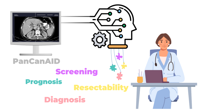
PanCanAID is committed to fostering research collaboration in academia and industry, in order to make use of AI-based early detection available for patients with a minimum cost. We are making our datasets and models available to qualified researchers, medical institutions, and device developers worldwide, with the aim of expediting progress in pancreatic cancer research and treatment modalities.
Notably, our license is the first of its kind to directly incorporate patient opinions regarding the use of their data. This patient-centric approach ensures that our data sharing practices align with the values and preferences of those most affected by pancreatic cancer.
Non-Profit Usage
- Eligible Entities: Academic researchers, medical centers, and device developers
- Requirements: Attribution of PanCanAID as the source of the dataset/model in all related publications and outputs
- Financial Considerations:
- No profit generation from the usage is permitted
- Users may charge patients to cover costs related to infrastructure and inference
- Any fees charged must be limited to cost recovery only
- Restrictions: Direct profit generation from our data or models is prohibited
Commercial Usage
- Eligible Entities: Commercial organizations developing applications or products
- Requirements:
- Allocation of 10% of profits to cancer-related charitable causes:
- Minimum 70% designated for patient assistance programs
- Maximum 30% allocated to AI development in cancer research
- Implementation Flexibility: We are open to discussing alternative arrangements, such as quinquennial (every 5 years) lump-sum payments, to accommodate various business models
Governance Structure
PanCanAID is committed to inclusive governance that represents key stakeholders. We are in the process of establishing a diverse board of directors, which will include:
- Three patient representatives
- The project founder, SAA Safavi-naini
- One representative from the development team
- One representative from the medical team
Board members will serve three-year terms. In the interim period before the board’s establishment, SAA Safavi-naini will serve as the primary governing authority for the project.
پروژه سرطان پانکراس (PanCanAID) متعهد به ترویج همکاریهای پژوهشی در میان دانشگاهها و صنایع است به گونه ای که با حداقل هزینه تحمیلی به بیماران، از منافع سیستم های مبتنی بر هوش مصنوعی بتوان بیماری را در مرحله زودرس تشخیص داد. ما مجموعههای دادهها و مدلهای خود را در اختیار پژوهشگران واجد شرایط، مؤسسات پزشکی و توسعهدهندگان دستگاههای پزشکی در سراسر جهان قرار میدهیم تا با هدف تسریع پیشرفت در پژوهشها و روشهای درمانی سرطان پانکراس به کار گرفته شوند.
قابل ذکر است که مجوز ما، اولین نمونهای است که نظرات بیماران در خصوص استفاده از دادههای آنها را به طور مستقیم لحاظ میکند. این رویکرد بیمار محور اطمینان میدهد که نحوه به اشتراک گذاری دادههای با ارزشها و ترجیحات افرادی که بیشترین تأثیر را از سرطان پانکراس میپذیرند، هماهنگ است.
استفاده غیرانتفاعی
نهادهای واجد شرایط: پژوهشگران دانشگاهی، مراکز پزشکی و توسعهدهندگان دستگاههای پزشکی
الزامات: ذکرPanCanAID به عنوان منبع مجموعه داده/مدل در تمام نشریات و خروجیهای مرتبط
ملاحظات مالی:
– کسب سود از استفاده از دادهها مجاز نیست.
– کاربران ممکن است برای پوشش هزینههای مربوط به زیرساخت و پیشبینی از بیماران هزینه دریافت کنند.
– هر گونه هزینهای که دریافت میشود باید محدود به جبران هزینهها باشد.
محدودیتها: کسب سود مستقیم از دادهها یا مدلهای ما ممنوع است تا هزینه استفاده از این ابزار ها برای بیماران به کمترین میزان ممکن برسد.
استفاده تجاری
نهادهای واجد شرایط: سازمانهای تجاری که در حال توسعه کاربردها یا محصولات هستند.
الزامات:
– تخصیص ۱۰٪ از سود به اهداف خیریه مرتبط با سرطان که شامل:
– حداقل ۷۰٪ به برنامههای کمک به بیماران اختصاص یابد.
– حداکثر ۳۰٪ به توسعه هوش مصنوعی در پژوهشهای سرطان اختصاص یابد.
انعطافپذیری در اجرا: ما آماده مذاکره در مورد ترتیبات جایگزین، مانند پرداختهای یکجا هر پنج سال یکبار، برای سازگاری با مدلهای تجاری مختلف هستیم.
ساختار اجرایی
ما در تلاش برای تشکیل هیئت تصمیم گیری هستیم که شامل موارد زیر خواهد بود:
– سه نماینده بیماران
– مؤسس پروژه، سید امیر احمد صفوی نائینی
– یک نماینده از تیم توسعه هوش مصنوعی
– یک نماینده از تیم پزشکی
اعضای هیئت صورت دورهای سه ساله خدمت خواهند کرد. در دوره موقت قبل از تشکیل این هیئت، سید امیر احمد صفوی نائینی به عنوان مرجع تصمیم گیری پروژه خدمت خواهد کرد.
PanCanAID Team
At our core, we believe that a culture of interdisciplinary collaboration and mutual respect is key to deploying AI successfully in healthcare practice. Our team is privileged to bring together senior experts and promising young researchers from diverse fields, all working together towards a common goal. With a positive culture that values each team member’s contributions and encourages ongoing learning, we’re able to tap into a wealth of collective experience and expertise to drive meaningful progress in healthcare innovation.

Seyed Amir Ahmad Safavi-Naini, MD-MBA
Research Fellow at RIGLD
Founder 🙂
Project Coordinators

Zahra Tajabadi
BS Student of Psychology at RIGLD

Elahe Meftah
MD student at TUMS

Elham Shabani
MD at MUMS
Model Development Team

Saeide Danaei
Bioinformatics MSc student at SUT

Zahra Dehghanian
PhD Candidate at SUT

Mehran Advand
Bioinformatics MSc student at SUT
Advisory Committee and Senior Members

Faezeh Khorasanizadeh, MD
Asst. Prof. Radiology at IKHC, TUMS

Amir Sadeghi, MD
Assoc. Prof. Gastroenterology at RIGLD, SBMU

Seyedmahdi Mirtajaddini, MD
Internist at TVH

Farhad Zamani, MD
Assoc. Prof. Gastroenterology at GILDRC, IUMS

Ali Soroush, MD-MSc
Asst. Prof. Gastroenterology at D3M Division, Icahn School of Medicine at Mount Sinai

Ashkan Zandi, PhD
Research Faculty at School of Electrical and Computer Engineering, Georgia Institute of Technology

Fatemeh Shojaeian, MD
Postdoc Fellow at Sidney Kimmel Comprehensive Cancer center, Johns Hopkins University

Pardis Ketabi Moghadam, MD
Asst. Prof. Gastroenterology at RIGLD, SBMU

Reza Farjad, MD
Asst. Prof. Radiology at SBMU and Milad Hospital
Reza Nafisi Moghadam, MD
Assoc. Prof. Radiology, Shahid Sadoughi University of Medical Sciences

Farahnaz Joukar, MSc, PhD
Assoc. Prof. Epidemiology at GLDRC
Research Assistant, Associate, and Fellow

Hamidreza Ghasemirad
Medical Sutdent at SSUMS

Mohammad Mohammadi
Medical Student at SSUMS

Abas Habibolahi
MD student at IUMS

Zahra Shahhoseini
Medical Student at SBMU

Parinaz Mellatdoust, MSc
Student at Dipartimento di elettronica informazione e bioingegneria, Politecnico di Milano

Alireza Mansour-Ghanaei, MD
Internal Medicine Resident at GUMS

Ghazale Sadeghi
Medical Student at SBMU

Aryan Salahy-Niri
BSc student Medical Labratory Science, SBMU

Mahsa Aghamohamadpor
Medical Student, SBMU

Mohammad Amin Tarighatpeima
Medical Student, SBMU
Server Maintenance

Maryam Saeedi, BSc
AI MSc Student at Islamic Azad University
Radiologists, Gastroenterologists, Surgeons, and Pathologists

Pooneh Dehghan
Assoc. Prof. Radiology at TH, SBMU

Faeze Salahshour, MD
Assoc. Prof. Radiology at IKHC, TUMS

Kavoos Firooznia, MD
Prof. Radiology at IKHC, TUMS

Shabnam Shahrokh, MD
Asst. Prof. Gastroentrology at RIGLD, SBMU

Masoomeh Raofi, MD
Assoc. Prof. Radiology at IHH, SBMU

Abdolhamid Chavoshi Khamneh, MD
Asst. Prof. Surgery at IUMS
Alireza Rasekhi, MD
Assoc. Prof. at SUMS

Zhaleh Mohsenifar, MD
Assoc. Prof. Pathology at SBMU

Akram Pourshams MD,MPH , MSc
Prof. Gastroenterology at DDRI, TUMS
Co-PI from DDRI

Farid Azmoudeh Ardalan, MD
Prof. Pathology at TUMS
MD Radiology Team

Elham Taghavi, MD
RIGLD

Sara Pourhemati, MD
RIGLD

Mahshad Sarikhani, MD

Benyamin Mohammadzadeh, MD

Ramin Shahidi, MD

Narges Azizi, MD
Research Associate
Radiologists Team

Mahyar Daskareh, MD

Azade Ehsani, MD

Mohamad Ghazanfari Hashemi, MD

Alireza salmanipour, MD

Faeze Shoja, MD
Senior Advisor

Mohammad Reza Zali, MD, FACG
Distinguished Professor of Gastroenterology and Hepatology at RIGLD, SBMU
Principial Investigator Core

Fariba Zarei, MD
Asst. Prof. Radiology at TUMS
Co-PI from SUMS

Fariborz Mansour-Ghanaei, MD, AGAF
Prof. Gastroentrology at GLDRC, GUMS
Co-PI from GUMS

Farzaneh Khoroushi, MD
َProf. Radiology at MUMS
Co-PI from MUMS

Masoudreza Sohrabi, MD-PhD
Senior Scientist at GILDRC, IUMS
Co-PI from IUMS

Hosein Ghanati, MD
Prof. Radiology at TUMS
Co-PI from TUMS

Hamid Assadzadeh Aghdaei, MD, PhD,
Assoc. Prof. Gastroenterology at RIGLD, SBMU
Co-PI and Medical Supervisor

Hamid R Rabiee, PhD
Distinguished Professor of Computer Science at DML, SUT
PI and ML Supervisor
This work has received support from multiple sources. The Research Institute for Gastroenterology and Liver Diseases, Shahid Beheshti University of Medical Sciences, provided funding for this project through Grant No. 0480/650 and Research No. 1224, which were granted to HAA and SAASN. HRR was partially supported by the IR National Science Foundation (INSF) through Grant No. 96006077. It is important to note that the funders did not and will not be involved in the study design, data collection, analysis, decision to publish, or manuscript preparation. Furthermore, SAASN provided additional funding for this project from personal funds in memory of Dr. Amanolah Safavi-Naini.

Email: sdamirsa@gmail.com Tel: +989120180209 Telegram: @PanCanAID